What Is Bursitis In The Foot?
Overview
A bursa is a fluid-containing sac that is present at many areas of pressure on the body. Its job is to protect these areas of pressure by being a shock absorber. A bursitis is an abnormal inflammation of the bursa sac caused by abnormal excessive pressure, stress and/ or strain over the area it is protecting. On the heel bone, there are two bursa sacs: one on the bottom and the other on the back. Specifically on the bottom of the heel, it is known as an Inferior (bottom) Calcaneal (heel) Bursa. On the back of the heel, it is called the Retro (back) Calcaneal Bursa. When either of these bursas become abnormally stressed, strained, or swollen, the result is bursitis of the heel. It is this bursitis that is the reason for pain in the heel upon arising (Poststatic Dyskinesia) in the morning or after resting for a while. You can either develop these bursitises with or without the presence of heel spur (explanation to follow). As stated before, Morton?s Toe can cause this by causing over pronation in the foot.
Causes
Overuse of the ankle joint may cause irritation of the bursa such as excessive walking, running or jumping. Poor biomechanics and foot function may ultimately lead to heel bursitis due to pulling on the back of the heel by the Achilles tendon.
A sudden increase in physical activity without adequate rest may result in heel bursitis. Excessive standing and walking bare foot on hard surfaces.
Symptoms
Symptoms of Achilles bursitis are often mistaken for Achilles tendinitis because of the location of the pain at the back of the heel. When you suffer from Achilles bursitis you will experience some or all of the following symptoms which are most noticeable when you begin activity after resting. High heels can add pressure on the retrocalcaneal bursa, subcutaneous calcaneal bursa, and Achilles tendon.
Diagnosis
Your doctor will examine you, including an evaluation of your gait, while you are barefoot, your doctor will ask you to stand still and to walk in order to evaluate how your foot moves as you walk. An examination of your feet. Your doctor may compare your feet for any differences between them. Then your doctor may examine your painful foot for signs of tenderness, swelling, discoloration, muscle weakness and decreased range of motion. A neurological examination. The nerves and muscles may be evaluated by checking strength, sensation and reflexes. In addition to examining you, your health care professional may want to examine your shoes. Signs of excessive wear in certain parts of a shoe can provide valuable clues to problems in the way you walk and poor bone alignment. Depending on the results of your physical examination, you may need foot X-rays or other diagnostic tests.
Non Surgical Treatment
Physical therapy is also used to treat retrocalcaneal bursitis. People with this condition may be instructed to use ice on the heel and ankle several times each day. Ice should be applied for periods of 15 to 20 minutes. Prolonged use of ice is not recommended because it can stop blood flow if left in place for a long period of time. Exercises and stretches for the Achilles tendon can help to relieve some of the pressure on the bursae below the tendons. If physical activity must be limited due to a flare-up of this condition, other exercises can be done to maintain fitness. They include water aerobics and swimming.
Surgical Treatment
Only if non-surgical attempts at treatment fail, will it make sense to consider surgery. Surgery for retrocalcanel bursitis can include many different procedures. Some of these include removal of the bursa, removing any excess bone at the back of the heel (calcaneal exostectomy), and occasionally detachment and re-attachment of the Achilles tendon. If the foot structure and shape of the heel bone is a primary cause of the bursitis, surgery to re-align the heel bone (calcaneal osteotomy) may be considered. Regardless of which exact surgery is planned, the goal is always to decrease pain and correct the deformity. The idea is to get you back to the activities that you really enjoy. Your foot and ankle surgeon will determine the exact surgical procedure that is most likely to correct the problem in your case. But if you have to have surgery, you can work together to develop a plan that will help assure success.
Prevention
People can lower the risk of bursitis by gradually strengthening and stretching the muscles around the joints and taking regular breaks from repetitive motion that might irritate bursae. Prolonged time resting on the elbows or kneeling should be avoided, if it cannot be avoided, wearing cushioned elbow and knee pads can help protect the bursae. Comfortable, supportive, low-heeled shoes can help prevent bursitis in the foot.
Hammer Toe Fusion Implants
 Overview
The 2nd toe is the most common digit to develop a hammertoes deformity. Second toe hammer toes commonly result from an elongated 2nd metatarsal and from pressure due to an excessively abducted great toe (hallux valgus deformity) causing a bunion. Unusually long toes often develop hammer toe deformities. Painful corns often develop in hammer toe deformity, particularly of the 5th toe. Reactive adventitial bursas often develop beneath corns, which may become inflamed.
Causes
A common cause of hammer toe is wearing shoes that do not fit properly. Poorly-fitting shoes can hold the toes in an abnormal position and result in tightening of the muscles required to maintain that position. In particular, shoes that have high heels and are narrow at front tend to push the toes into an abnormal, bent position. Less commonly, diseases of the nerves, muscles, or joints (such as arthritis) can result in the hammer toe deformity.
 Symptoms
Signs and symptoms of hammertoe and mallet toe may include a hammer-like or claw-like appearance of a toe. In mallet toe, a deformity at the end of the toe, giving the toe a mallet-like appearance. Pain and difficulty moving the toe. Corns and calluses resulting from the toe rubbing against the inside of your footwear. Both hammertoe and mallet toe can cause pain with walking and other foot movements.
Diagnosis
Hammer toes may be easily detected through observation. The malformation of the person's toes begin as mild distortions, yet may worsen over time - especially if the factors causing the hammer toes are not eased or removed. If the condition is paid attention to early enough, the person's toes may not be permanently damaged and may be treated without having to receive surgical intervention. If the person's toes remain untreated for too long, however the muscles within the toes might stiffen even more and will require invasive procedures to correct the deformity.
Non Surgical Treatment
If the problem is caught in the early stages you can avoid hammer toe surgery. One of the easiest methods of treatment is to manipulate the toe out of a bent position then splint and buddy wrap it alongside it?s larger neighbour. This method of hammer toe taping will help the problem to fix itself. Make sure the toe isn?t resuming its bent shape during the recovery. To alleviate some of the painful symptoms of hammer toe avoid wearing high heels or shoes that cramp or stifle your feet. Choosing a pair of minimalist shoes can be an excellent choice for both foot and postural health. Wearing shoes that give the toes plenty of hammertoes space and are comfortable lined is also a smart choice. Hammer toe recovery starts be treating the toe respectfully. Soft insoles or protection for the corn can also provide additional assistance.
Surgical Treatment
The deformity is corrected in a variety of ways. There are actually a large number of procedures. The simplest procedure would involve a Tenotomy, the cutting of the tendon causing the deformity or a Tendon Lengthening procedure. These procedures are infrequently done, though, as the structural deformity (the arthritis and joint adaptation) is not addressed with these surgeries. Other soft-tissue procedures involve rebalancing the tendons around the joint. There are several techniques to do this, but the most common is probably the Girdlestone-Taylor procedure, which involves rerouting the tendons on the bottom of the toe up and over the toe where it sticks up, so that the tendon helps pull the toe downwards into proper alignment.
The Truth Regarding Over-Pronation
Overview
Your feet are the foundation for your entire body. When this foundation is misaligned or functioning poorly the effects can be felt throughout the body, whether in muscle and joint pain or through more serious injuries. Over 75% of the population suffers from overpronation or excessive supination, yet most of us are unaware of our own foot type and how it affects the rest of our body.
Causes
Over-pronation may happen because the tissue that attaches to your foot bones is loose. You may be born with this problem or it may result from injuries or overuse, like from too much running.
Symptoms
Because pronation is a twisting of the foot, all of the muscles and tendons which run from the leg and ankle into the foot will be twisted. In over-pronation, resulting laxity of the soft tissue structures of the foot and loosened joints cause the bones of the feet shift. When this occurs, the muscles which attach to these bones must also shift, or twist, in order to attach to these bones. The strongest and most important muscles that attach to our foot bones come from our lower leg. So, as these muscles course down the leg and across the ankle, they must twist to maintain their proper attachments in the foot. Injuries due to poor biomechanics and twisting of these muscles due to over-pronation include: shin splints, Achilles Tendonitis, generalized tendonitis, fatigue, muscle aches and pains, cramps, ankle sprains, and loss of muscular efficiency (reducing walking and running speed and endurance). Foot problems due to over-pronation include: bunions, heel spurs, plantar fasciitis, fallen and painful arches, hammer toes, and calluses.
Diagnosis
When you overpronate your foot rolls inwards causing the lower leg to rotate inwards too. It's thought that this increases stress on the soft tissues of the lower leg causing pain and inflammation, or more commonly known as shin splints.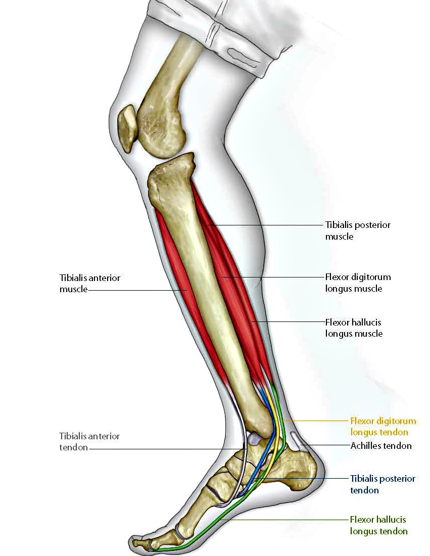
Non Surgical Treatment
Supportive orthotics in the shoe is a method commonly implemented to treat many common running injuries associated with pronation. An advantage of orthotics is that they often allow the sufferer to continue to participate in athletic activity and avoid other treatment options that could be potentially costly and time consuming. Seventy-five percent of injured runners are successfully treated with the prescription of orthoses. Orthotics are the most effective treatment for symptoms that develop from unusual biomechanics within the body such as overpronation, resulting in either great improvement or complete healing of the injury in about half the cases.
Surgical Treatment
Hyperpronation can only be properly corrected by internally stabilizing the ankle bone on the hindfoot bones. Several options are available. Extra-Osseous TaloTarsal Stabilization (EOTTS) There are two types of EOTTS procedures. Both are minimally invasive with no cutting or screwing into bone, and therefore have relatively short recovery times. Both are fully reversible should complications arise, such as intolerance to the correction or prolonged pain. However, the risks/benefits and potential candidates vary. Subtalar Arthroereisis. An implant is pushed into the foot to block the excessive motion of the ankle bone. Generally only used in pediatric patients and in combination with other procedures, such as tendon lengthening. Reported removal rates vary from 38% - 100%, depending on manufacturer. HyProCure Implant. A stent is placed into a naturally occurring space between the ankle bone and the heel bone/midfoot bone. The stent realigns the surfaces of the bones, allowing normal joint function. Generally tolerated in both pediatric and adult patients, with or without adjunct soft tissue procedures. Reported removal rates, published in scientific journals vary from 1%-6%.
Diagnosing Severs Disease
Overview
When recurring heel pain occurs in children, it is usually due to Sever's Disease, while adult heel pain is usually due to heel spurs, plantar fasciitis, or retrocalcaneal bursitis (Haglund's Deformity). Calcaneus is the anatomical name of the heel bone. Sever's Disease or Calcaneal Apophysitis is an inflammation of the growth plate located at the posterior aspect (back) of the heel.
Causes
Severs disease is often associated with a rapid growth spurt. As the bones get longer, the muscles and tendons become tighter as they cannot keep up with the bone growth. The point at which the achilles tendon attaches to the heel becomes inflamed and the bone starts to crumble (a lot like osgood schlatters disease of the knee). Tight calf muscles may contribute as the range of motion at the ankle is reduced resulting in more strain on the achilles tendon. Sever's disease is the second most common injury of this type which is known as an apophysitis.
Symptoms
Sever condition causes pain at the back of the heel. The pain is increased with plantar flexion of the ankle (pushing down with the foot as if stepping on the gas), particularly against resistance. Sever condition also causes tenderness and swelling in the area of the pain.
Diagnosis
A doctor can usually tell that a child has Sever's disease based on the symptoms reported. To confirm the diagnosis, the doctor will probably examine the heels and ask about the child's activity level and participation in sports. The doctor might also use the squeeze test, squeezing the back part of the heel from both sides at the same time to see if doing so causes pain. The doctor might also ask the child to stand on tiptoes to see if that position causes pain. Although imaging tests such as X-rays generally are not that helpful in diagnosing Sever's disease, some doctors order them to rule out other problems, such as fractures. Sever's disease cannot be seen on an X-ray.
Non Surgical Treatment
Your podiatrist can help manage this condition by implementing a treatment program. This may incorporate one or all of the following. RI (Rest and Ice). Activity modification so child becomes pain free. Daily stretching routine. Heel raise within shoes to decrease pull on heel. Biomechanical abnormalities corrected (Orthotics). Strengthening of associated muscles. Footwear modification.
Recovery
It may take several weeks or months for the pain to completely stop. In most cases severs disease goes away on its own with a little rest and time. However if you ignore the pain and play through it, the condition may get worse and may be more difficult to treat. When the pain is completely gone, you can slowly return to your previous level of activity. With future growth spurts the pain may return therefore keep up with the stretches and follow the advice given.
Acquired Flatfoot Deformity
Overview
PTTD is a common condition treated by foot and ankle specialists. Although there is a role for surgical treatment of PTTD, conservative care often can prevent or delay surgical intervention. Decreasing inflammation and stabilizing the affected joints associated with the posterior tibial tendon can decrease pain and increase functional levels. With many different modalities available, aggressive nonoperative methods should be considered in the treatment of PTTD, including early immobilization, the use of long-term bracing, physical therapy, and anti-inflammatory medications. If these methods fail, proper evaluation and work-up for surgical intervention should be employed. 
Causes
Adult flatfoot typically occurs very gradually. If often develops in an obese person who already has somewhat flat feet. As the person ages, the tendons and ligaments that support the foot begin to lose their strength and elasticity.
Symptoms
The symptom most often associated with AAF is PTTD, but it is important to see this only as a single step along a broader continuum. The most important function of the PT tendon is to work in synergy with the peroneus longus to stabilize the midtarsal joint (MTJ). When the PT muscle contracts and acts concentrically, it inverts the foot, thereby raising the medial arch. When stretched under tension, acting eccentrically, its function can be seen as a pronation retarder. The integrity of the PT tendon and muscle is crucial to the proper function of the foot, but it is far from the lone actor in maintaining the arch. There is a vital codependence on a host of other muscles and ligaments that when disrupted leads to an almost predictable loss in foot architecture and subsequent pathology.
Diagnosis
The diagnosis of tibialis posterior dysfunction is essentially clinical. However, plain radiographs of the foot and ankle are useful for assessing the degree of deformity and to confirm the presence or absence of degenerative changes in the subtalar and ankle articulations. The radiographs are also useful to exclude other causes of an acquired flatfoot deformity. The most useful radiographs are bilateral anteroposterior and lateral radiographs of the foot and a mortise (true anteroposterior) view of the ankle. All radiographs should be done with the patient standing. In most cases we see no role for magnetic resonance imaging or ultrasonography, as the diagnosis can be made clinically.
Non surgical Treatment
Conservative treatment is indicated for nearly all patients initially before surgical management is considered. The key factors in determining appropriate treatment are whether acute inflammation and whether the foot deformity is flexible or fixed. However, the ultimate treatment is often determined by the patients, most of whom are women aged 40 or older. Compliance can be a problem, especially in stages I and II. It helps to emphasise to the patients that tibialis posterior dysfunction is a progressive and chronic condition and that several fittings and a trial of several different orthoses or treatments are often needed before a tolerable treatment is found. 
Surgical Treatment
Surgery is usually performed when non-surgical measures have failed. The goal of surgery is to eliminate pain, stop progression of the deformity and improve a patient?s mobility. More than one technique may be used, and surgery tends to include one or more of the following. The tendon is reconstructed or replaced using another tendon in the foot or ankle The name of the technique depends on the tendon used. Flexor digitorum longus (FDL) transfer. Flexor hallucis longus (FHL) transfer. Tibialis anterior transfer (Cobb procedure). Calcaneal osteotomy - the heel bone may be shifted to bring your heel back under your leg and the position fixed with a screw. Lengthening of the Achilles tendon if it is particularly tight. Repair one of the ligaments under your foot. If you smoke, your surgeon may refuse to operate unless you can refrain from smoking before and during the healing phase of your procedure. Research has proven that smoking delays bone healing significantly.
What Is Adult Aquired FlatFeet ?
Overview
Adult flatfoot (adult acquired flatfoot) or posterior tibial tendon dysfunction (PTTD) is a common pathology presented to foot and ankle specialists. PTTD is characterized by a valgus (everted) hindfoot, flattening of the longitudinal arch of the foot (collapse) and abduction of the forefoot. This is a progressive deformity that begins flexible and can become rigid over time. The posterior tibial tendon (PT) is one of the main supporting structures of the foot arch. Changes within this tendon cause flattening of the foot. There are four stages of this deformity that begins flexible and progressives, with no treatment, to a rigid deformity and with time may involve the ankle joint. Patients usually present with pain in the foot or ankle stating the ankle is rolling. Its also common for patients to state they have difficulty walking barefoot. Pain is exacerbated after physical activities. Pain is usually isolated to the inside of the foot along the course of the PT tendon.
Causes
Many health conditions can create a painful flatfoot, an injury to the ligaments in the foot can cause the joints to fall out of alignment. The ligaments support the bones and prevent them from moving. If the ligaments are torn, the foot will become flat and painful. This more commonly occurs in the middle of the foot (Lisfranc injury), but can also occur in the back of the foot. In addition to ligament injuries, fractures and dislocations of the bones in the midfoot can also lead to a flatfoot deformity.
Symptoms
The symptom most often associated with AAF is PTTD, but it is important to see this only as a single step along a broader continuum. The most important function of the PT tendon is to work in synergy with the peroneus longus to stabilize the midtarsal joint (MTJ). When the PT muscle contracts and acts concentrically, it inverts the foot, thereby raising the medial arch. When stretched under tension, acting eccentrically, its function can be seen as a pronation retarder. The integrity of the PT tendon and muscle is crucial to the proper function of the foot, but it is far from the lone actor in maintaining the arch. There is a vital codependence on a host of other muscles and ligaments that when disrupted leads to an almost predictable loss in foot architecture and subsequent pathology.
Diagnosis
The diagnosis of posterior tibial tendon dysfunction and AAFD is usually made from a combination of symptoms, physical exam and x-ray imaging. The location of pain, shape of the foot, flexibility of the hindfoot joints and gait all may help your physician make the diagnosis and also assess how advanced the problem is.
Non surgical Treatment
It is imperative that you seek treatment should you notice any symptoms of a falling arch or PTTD. Due to the progressive nature of this condition, your foot will have a much higher chance of staying strong and healthy with early treatment. When pain first appears, your doctor will evaluate your foot to confirm a flatfoot diagnosis and begin an appropriate treatment plan. This may involve rest, anti-inflammatory medications, shoe modifications, physical therapy, orthotics and a possible boot or brace. When treatment can be applied at the beginning, symptoms can most often be resolved without the need for surgery.

Surgical Treatment
The indications for surgery are persistent pain and/or significant deformity. Sometimes the foot just feels weak and the assessment of deformity is best done by a foot and ankle specialist. If surgery is appropriate, a combination of soft tissue and bony procedures may be considered to correct alignment and support the medial arch, taking strain off failing ligaments. Depending upon the tissues involved and extent of deformity, the foot and ankle specialist will determine the necessary combination of procedures. Surgical procedures may include a medial slide calcaneal osteotomy to correct position of the heel, a lateral column lengthening to correct position in the midfoot and a medial cuneiform osteotomy or first metatarsal-tarsal fusion to correct elevation of the medial forefoot. The posterior tibial tendon may be reconstructed with a tendon transfer. In severe cases (stage III), the reconstruction may include fusion of the hind foot,, resulting in stiffness of the hind foot but the desired pain relief. In the most severe stage (stage IV), the deltoid ligament on the inside of the ankle fails, resulting in the deformity in the ankle. This deformity over time can result in arthritis in the ankle.
Adult flatfoot (adult acquired flatfoot) or posterior tibial tendon dysfunction (PTTD) is a common pathology presented to foot and ankle specialists. PTTD is characterized by a valgus (everted) hindfoot, flattening of the longitudinal arch of the foot (collapse) and abduction of the forefoot. This is a progressive deformity that begins flexible and can become rigid over time. The posterior tibial tendon (PT) is one of the main supporting structures of the foot arch. Changes within this tendon cause flattening of the foot. There are four stages of this deformity that begins flexible and progressives, with no treatment, to a rigid deformity and with time may involve the ankle joint. Patients usually present with pain in the foot or ankle stating the ankle is rolling. Its also common for patients to state they have difficulty walking barefoot. Pain is exacerbated after physical activities. Pain is usually isolated to the inside of the foot along the course of the PT tendon.
Causes
Many health conditions can create a painful flatfoot, an injury to the ligaments in the foot can cause the joints to fall out of alignment. The ligaments support the bones and prevent them from moving. If the ligaments are torn, the foot will become flat and painful. This more commonly occurs in the middle of the foot (Lisfranc injury), but can also occur in the back of the foot. In addition to ligament injuries, fractures and dislocations of the bones in the midfoot can also lead to a flatfoot deformity.
Symptoms
The symptom most often associated with AAF is PTTD, but it is important to see this only as a single step along a broader continuum. The most important function of the PT tendon is to work in synergy with the peroneus longus to stabilize the midtarsal joint (MTJ). When the PT muscle contracts and acts concentrically, it inverts the foot, thereby raising the medial arch. When stretched under tension, acting eccentrically, its function can be seen as a pronation retarder. The integrity of the PT tendon and muscle is crucial to the proper function of the foot, but it is far from the lone actor in maintaining the arch. There is a vital codependence on a host of other muscles and ligaments that when disrupted leads to an almost predictable loss in foot architecture and subsequent pathology.
Diagnosis
The diagnosis of posterior tibial tendon dysfunction and AAFD is usually made from a combination of symptoms, physical exam and x-ray imaging. The location of pain, shape of the foot, flexibility of the hindfoot joints and gait all may help your physician make the diagnosis and also assess how advanced the problem is.
Non surgical Treatment
It is imperative that you seek treatment should you notice any symptoms of a falling arch or PTTD. Due to the progressive nature of this condition, your foot will have a much higher chance of staying strong and healthy with early treatment. When pain first appears, your doctor will evaluate your foot to confirm a flatfoot diagnosis and begin an appropriate treatment plan. This may involve rest, anti-inflammatory medications, shoe modifications, physical therapy, orthotics and a possible boot or brace. When treatment can be applied at the beginning, symptoms can most often be resolved without the need for surgery.
Surgical Treatment
The indications for surgery are persistent pain and/or significant deformity. Sometimes the foot just feels weak and the assessment of deformity is best done by a foot and ankle specialist. If surgery is appropriate, a combination of soft tissue and bony procedures may be considered to correct alignment and support the medial arch, taking strain off failing ligaments. Depending upon the tissues involved and extent of deformity, the foot and ankle specialist will determine the necessary combination of procedures. Surgical procedures may include a medial slide calcaneal osteotomy to correct position of the heel, a lateral column lengthening to correct position in the midfoot and a medial cuneiform osteotomy or first metatarsal-tarsal fusion to correct elevation of the medial forefoot. The posterior tibial tendon may be reconstructed with a tendon transfer. In severe cases (stage III), the reconstruction may include fusion of the hind foot,, resulting in stiffness of the hind foot but the desired pain relief. In the most severe stage (stage IV), the deltoid ligament on the inside of the ankle fails, resulting in the deformity in the ankle. This deformity over time can result in arthritis in the ankle.
Coping With Achilles Tendinitis Problems
Overview
 Achilles Tendinitis is the inflammation of the Achilles Tendon located in the heel, and is typically caused by overuse of the affected limb. Most often, it occurs in athletes who are not training with the proper techniques and/or equipment. When the Achilles Tendon is injured, blood vessels and nerve fibers from surrounding areas migrate into the tendon, and the nerve fibers may be responsible for the discomfort. Healing is often slow in this area due to the comparably low amount of cellular activity and blood flowing through the area.
Causes
Achilles tendinitis may be caused by intensive hill running, sprinting, or stair climbing. Overuse resulting from the natural lack of flexibility in the calf muscles. Rapidly increasing intensity of exercise, especially after a period of inactivity. Sudden and hard contraction of the calf muscles when exerting extra effort, like that in a final sprint or high jump.
Symptoms
Mild ache in the back of the lower leg, especially after running. More acute pain may occur after prolonged activity, Tenderness or stiffness in the morning. In most cases the pain associated with Achilles tendinitis is more annoying than debilitating, making sufferers regret activity after the fact, but not keeping them from doing it. More severe pain around the Achilles tendon may be a symptom of a much more serious ruptured tendon.
Diagnosis
If you think you have Achilles tendinitis, make an appointment to see your doctor. The doctor will ask you questions about your recent activity and look for signs. The foot not flexing when the calf muscle is pressed ( if Achilles ruptures or tears in half). Swelling on the back of the foot. Pain in the back of the foot. Limited range of motion in ankle. An X-ray or MRI scan can check for tendinitis.
Nonsurgical Treatment
Use the R.I.C.E method of treatment when you first notice the pain. Although rest is a key part of treating tendonitis, prolonged inactivity can cause stiffness in your joints. Move the injured ankle through its full range of motion and perform gentle calf and ankle stretches to maintain flexibility. If self-care doesn't work, it's important to get the injury treated because if the tendon continues to sustain small tears through movement, it can rupture under excessive stress. Your doctor may suggest a temporary foot insert that elevates your heel and may relieve strain on the tendon. Other possible treatments include special heel pads or cups to wear in your shoes to cushion and support your heel, or a splint to wear at night. Physical therapy may also help allow the tendon to heal and repair itself over a period of weeks.
Surgical Treatment
Occasionally, conservative management of Achilles tendon conditions fails. This failure is more common in older male patients and those with longstanding symptoms, those who persist in full training despite symptoms or those who have uncorrected predisposing factors. In these cases, surgery may be indicated. It should be remembered, however, that the rehabilitation program, particularly for severe Achilles tendon injuries, is a slow, lengthy program. Surgery is only indicated when there is failure to progress in the rehabilitation program. Surgery should not be considered unless at least six months of appropriate conservative management has failed to lead to improvement.
Prevention
Wear shoes that fit correctly and support your feet: Replace your running or exercise shoes before the padding or shock absorption wears out. Shock absorption greatly decreases as the treads on the bottoms or sides of your shoes begin to wear down. You may need running shoes that give your foot more heel or arch support. You may need shoe inserts to keep your foot from rolling inward. Stretch before you exercise: Always warm up your muscles and stretch gently before you exercise. Do cool down exercises when you are finished. This will loosen your muscles and decrease stress on your Achilles tendon. Exercise the right way: If your tendinitis is caused by the way that you exercise, ask a trainer, coach, or your caregiver for help. They can teach you ways to train or exercise to help prevent Achilles tendinitis. Do not run or exercise on uneven or hard surfaces. Instead, run on softer surfaces such as treadmills, rubber tracks, grass, or evenly packed dirt tracks.
Achilles Tendinitis is the inflammation of the Achilles Tendon located in the heel, and is typically caused by overuse of the affected limb. Most often, it occurs in athletes who are not training with the proper techniques and/or equipment. When the Achilles Tendon is injured, blood vessels and nerve fibers from surrounding areas migrate into the tendon, and the nerve fibers may be responsible for the discomfort. Healing is often slow in this area due to the comparably low amount of cellular activity and blood flowing through the area.
Causes
Achilles tendinitis may be caused by intensive hill running, sprinting, or stair climbing. Overuse resulting from the natural lack of flexibility in the calf muscles. Rapidly increasing intensity of exercise, especially after a period of inactivity. Sudden and hard contraction of the calf muscles when exerting extra effort, like that in a final sprint or high jump.
Symptoms
Mild ache in the back of the lower leg, especially after running. More acute pain may occur after prolonged activity, Tenderness or stiffness in the morning. In most cases the pain associated with Achilles tendinitis is more annoying than debilitating, making sufferers regret activity after the fact, but not keeping them from doing it. More severe pain around the Achilles tendon may be a symptom of a much more serious ruptured tendon.
Diagnosis
If you think you have Achilles tendinitis, make an appointment to see your doctor. The doctor will ask you questions about your recent activity and look for signs. The foot not flexing when the calf muscle is pressed ( if Achilles ruptures or tears in half). Swelling on the back of the foot. Pain in the back of the foot. Limited range of motion in ankle. An X-ray or MRI scan can check for tendinitis.
Nonsurgical Treatment
Use the R.I.C.E method of treatment when you first notice the pain. Although rest is a key part of treating tendonitis, prolonged inactivity can cause stiffness in your joints. Move the injured ankle through its full range of motion and perform gentle calf and ankle stretches to maintain flexibility. If self-care doesn't work, it's important to get the injury treated because if the tendon continues to sustain small tears through movement, it can rupture under excessive stress. Your doctor may suggest a temporary foot insert that elevates your heel and may relieve strain on the tendon. Other possible treatments include special heel pads or cups to wear in your shoes to cushion and support your heel, or a splint to wear at night. Physical therapy may also help allow the tendon to heal and repair itself over a period of weeks.
Surgical Treatment
Occasionally, conservative management of Achilles tendon conditions fails. This failure is more common in older male patients and those with longstanding symptoms, those who persist in full training despite symptoms or those who have uncorrected predisposing factors. In these cases, surgery may be indicated. It should be remembered, however, that the rehabilitation program, particularly for severe Achilles tendon injuries, is a slow, lengthy program. Surgery is only indicated when there is failure to progress in the rehabilitation program. Surgery should not be considered unless at least six months of appropriate conservative management has failed to lead to improvement.
Prevention
Wear shoes that fit correctly and support your feet: Replace your running or exercise shoes before the padding or shock absorption wears out. Shock absorption greatly decreases as the treads on the bottoms or sides of your shoes begin to wear down. You may need running shoes that give your foot more heel or arch support. You may need shoe inserts to keep your foot from rolling inward. Stretch before you exercise: Always warm up your muscles and stretch gently before you exercise. Do cool down exercises when you are finished. This will loosen your muscles and decrease stress on your Achilles tendon. Exercise the right way: If your tendinitis is caused by the way that you exercise, ask a trainer, coach, or your caregiver for help. They can teach you ways to train or exercise to help prevent Achilles tendinitis. Do not run or exercise on uneven or hard surfaces. Instead, run on softer surfaces such as treadmills, rubber tracks, grass, or evenly packed dirt tracks.